148
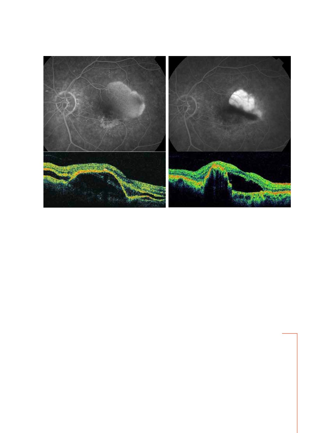
Figure 5 - Vascularized PED with choroidal neovascularization before and after PDT: RPE tear (fluorescein angiography and OCT)
A
B
and scanty response to the treatment frequently invali-
date our attempts to heal the lesion. RPE tears and sub-
retinal hemorrhages are reported to complicate ranibi-
zumab and bevacizumab treatments
(51-57)
. Moreover, the
sub-RPE fluid hardly responds to the anti-VEGF ther-
apy, probably due to the hydroconductivity changes of
Bruch’s membrane
(68)
. In a recent retrospective case series
of 328 patients treated with bevacizumab, ranibizumab,
pegaptanib and PDT+IVTA respectively, after a mean
follow-up of 42, 4 weeks the authors reported a significant
stabilization of visual acuity in each group, better in the
bevacizumab and ranibizumab ones compared to the
other two, and an overall RPE tears frequency of 12,5%.
However, they conclude that with these treatments, only
a partial regression of the lesions can be obtained, and
the risk of RPE tears is not avoided
(68)
. In the future, new
combination therapies and new therapeutic strategies,
tested in multicentric clinical trials specifically designed,
will help to improve the prognosis of the patients affected
by vascularized PED.
Correspondence concerning this article can be sent directly to the author through the email:


