63
3. Angiographic patterns in AMD
3.1 Drusen and RPE abnormalities
The majority of patients with AMD have drusen and RPE
abnormalities with no significant visual loss. FA is not usu-
ally indicated in these cases unless we suspect the presence
of choroidal neovascularization (CNV). Several types of
drusen can be identified. Hard drusen are small (<63 µm),
round, well-defined, yellowish deposits that correspond to
accumulation of hyaline material in the inner and outer
collagenous zones of Bruch’s membrane. On FA, they
appear hyperfluorescent as transmission defects due to
overlying RPE thinning
(6)
. On occasion there may be a
myriad of small drusen, termed cuticular or basal laminar
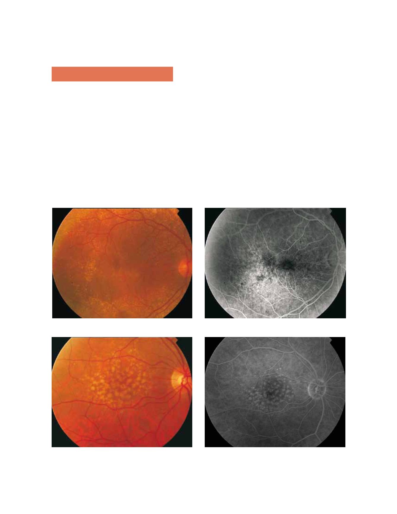
drusen, which appear as a “starry sky” on FA (Fig. 1). Soft
drusen are larger (>63 µm) with poorly defined borders
and they tend to coalesce and become confluent. Their
angiographic appearance depends on the thinning of the
overlying RPE, the histochemical composition and the
age of the patient. They are hyperfluorescent with phos-
pholipid accumulation and in younger patients
(7)
. Soft
drusen represent localized detachments of the RPE. It is
very usual to find both hard and soft drusen in the same
eye of a patient (Fig. 2). The confluence of soft drusen
can produce a drusenoid pigment epithelial detachment
(PED), which shows hyperfluorescence and dye pooling
without leakage beyond its margin with typical areas of
focal hyperpigmentation (Fig. 3).
In addition to drusen we can find RPE abnormalities,
namely hyperpigmentation. Focal hyperpigmentation is
a risk factor for the development of choroidal neovas-
cularization (CNV) and angiographically appears as a
Figure 1 - Cuticular drusen with the typical pattern of “starry sky”
Figure 2 - Coexistence of hard and soft drusen in the same eye of a patient with AMD.
Fluorescein Angiography


