66
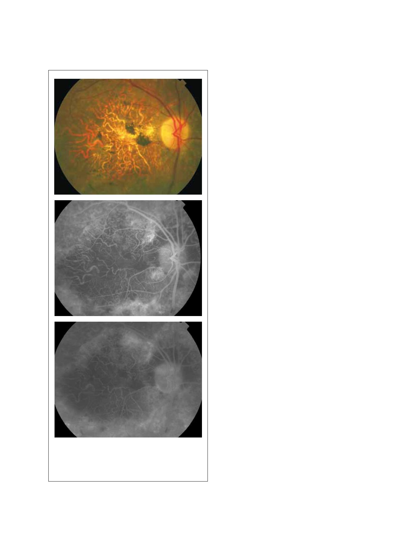
Figure 7 - Severe atrophic AMD with sclerotic appearance of larger
choroidal vessels.
blocked fluorescence (Fig. 4). Histopathologically it is
characterized by focal RPE hypertrophy and pigment
migration into the subretinal space. It also displays focal
hyperautofluorescence suggesting that these cells contain
lipofuscin
(8)
.
3.2 Atrophic AMD
Atrophy can occur in sharply defined areas of severe atro-
phy, known as geographic atrophy (GA), or in less well-
defined, more granular regions of less severe atrophy,
known as non-GA. Both forms share the feature of RPE
loss, more extensive and with associated atrophy of the
overlying retina and underlying choriocapillaris in GA.
The angiographic appearance depends on the remain-
ing pigment within the RPE and choriocapillaris vessels.
Non-GA shows mottled early hyperfluorescence, which
fades late consistent with window defect (Fig. 5). GA
typically shows late well-defined hyperfluorescence from
staining of the exposed deep choroid and sclera
(9)
. In
these cases, visual acuity depends on the foveal involve-
ment (Fig. 6). In advanced cases, larger choroidal vessels
show a sclerotic appearance (Fig. 7).
3.3 Classic CNV
Classic CNV is characterized by well-demarcated hyper-
fluorescence in early phases on FA and late leakage that
obscures the boundaries of the lesion (Fig. 8). As defined
by Donald Gass, classic CNV lies between the neurosen-
sory retina and the RPE (type II CNV)
(10)
. Angiographic
classic CNV appears as a lacy or bicycle-wheel pattern.
Depending on its location, it can be classified as extrafo-
veal (>200 microns from the foveal center) (Fig. 9), jux-
tafoveal (1-199 microns from the foveal center) (Fig. 10)
or subfoveal (involving the foveal center) (Fig. 11).
Sometimes, a feeder vessel can be localized (Fig. 12).
Another typical feature is the presence of a hyperpig-
mented rim, hypofluorescent on FA, surrounding the
CNV (Fig. 12). On occasion, classic CNV can be associ-
ated to loculated fluid (Fig. 13). In loculated fluid, dye
pooling is well-demarcated in a confined space of a local-
ized sensory retinal detachment or within intraretinal
cystic spaces. It was a common finding in patients with
new subfoveal CNV in the Macular Photocoagulation
Study (MPS) and may confuse the treating physician
as to the boundary of the lesion
(11)
. Depending on their
sizes, classic CNV can be classified as small (Fig. 9-11) or
medium (Fig. 14) or large (Fig. 15).


