132
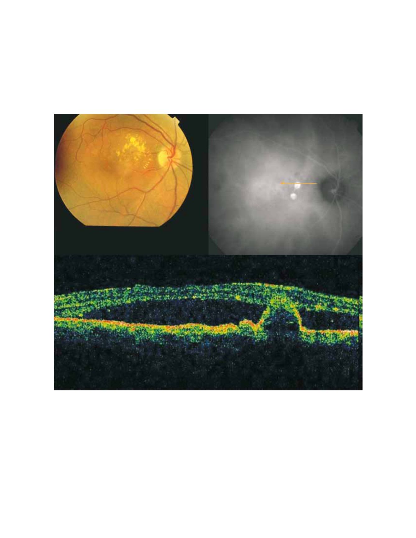
Figure 2- Fundus colour photography with lipidic exudation and a redish-orange, spheroid, subretinal mass located at the macular area associated
with macular edema. Late ICG (top, right) reveals the presence of two polypoidal lesions in the papilomacular bundle. On OCT (bottom) the polyp
is well delineated and a sub-foveal neurosensory detachment is observed.


