122
focal hyperreflective area located in the deep retina (intra-
retinal neovascularization), as well as being often associ-
ated with intraretinal fluid with cystic spaces, subretinal
fluid and PED (Fig. 4). In stage III lesions, neovascular
proliferation associated with PED may be observed.
With time-domain OCT
(20,21)
, (TD-OCT) a typical pat-
tern of structural changes in RAP may be observed, char-
acterized by increased foveal thickness, cystoid macular
oedema (CME) mainly located in outer retinal layers,
serous retinal detachment and a highly reflective intrareti-
nal mass overlying a highly or moderately elevated retinal
pigment epithelium. This mass corresponds to the hot
spot observed in ICG angiography. With Fourier-domain
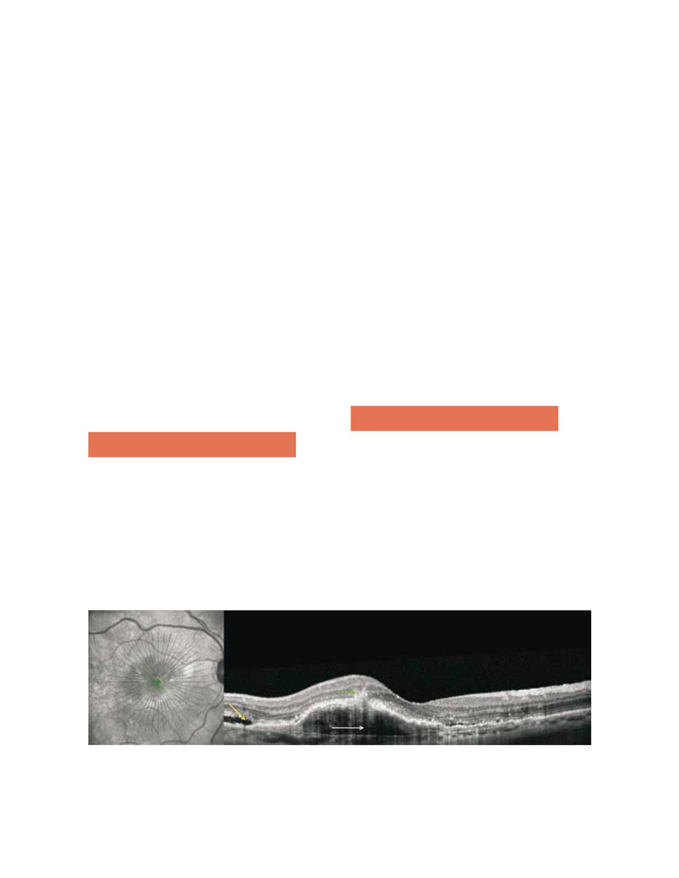
OCT (TD-OCT) it is possible to obtain unprecedented
in vivo
detail of the anatomy of RAP lesions, with images
nearly resembling histological specimens. OCT findings
may vary with the stage of the disease and type of early
neovascularization, including
(22,23)
areas of intraretinal
neovascularization (IRN) in the deep retina, adjacent to
PED, anterior and posterior neovascular proliferation
through a break in the RPE, intact and ruptured portions
of Bruch’s membrane, subretinal fluid and subretinal and/
or sub-RPE neovascular membranes (Fig. 5).
4. Differential diagnosis
Differential diagnosis is mandatory for parafoveal telangi-
ectasias, other forms of choroidal neovascularization and
polypoidal choroidal vasculopathy.
Idiopathic parafoveal telangiectasia is a condition involv-
ing dilation of retinal capillaries located near the fovea, in
one or both eyes. RPE hyperplasia may also occur, with
refractive punctiform deposits and macular leakage being
observed in FA. Migration of one or more venules to the
deep retina may also be observed
(5)
. Anastomoses between
retinal vessels and the choroidal circulation have been
described, as well as new choroidal vessels. The most sig-
nificant differences are the fact that telangiectasias are not
associated with serous PED, the RPE is healthier and cho-
roidal neovascularization associated with parafoveal telan-
giectasias occurs less frequently
(5,10)
.
Differential diagnosis should also be performed for other
forms of choroidal neovascularization (CNV) with ICG
hot spots (occult CNV) and polypoidal choroidal vasculop-
athy (PCV). Small intraretinal haemorrhages, sometimes
punctiform, in patients with soft drusen, are very typical
in RAP, as are telangiectasias and retino-retinal anastomo-
ses. Retinal haemorrhages in PCV are normally larger, with
round reddish-orange macular lesions being observed in
the eye fundus. OCT is also a useful differential diagnosis
tool in RAP, PCV and occult membranes. In RAP, intra-
retinal hyperreflectivity may be observed, corresponding to
angiomatous proliferation associated with intraretinal fluid
and/or RPE detachment. In PCV, polyps appear in OCT
as abrupt protrusions from the REP/Bruch’s membrane
band, often associated with neurosensory detachment.
5. Natural progression
Natural history may be highly variable and probably simi-
lar to that of other CNV lesions. However, many reports
ascribe a poor prognosis to RAP lesions. Kuhn et al.
(12)
studied 22 eyes and observed structural evolution towards
classic membranes, signs of RPE rupture and fibrous scars
in 36.4%, 4,6% and 31,8% of cases, respectively. In func-
tional terms, decrease in visual acuity occurred in 77%
of cases. Final visual acuity (VA) values equal or inferior
to 20/200 were observed in 14 eyes studied by Hartnett
et al.
(17)
. In a retrospective and one-year study performed
by Silva et al.
(14)
in 17 consecutive patients with RAP, a
Figure 5 - RAP lesion with an area of intraretinal neovascularization (green arrow) in the deep retina with pigment epithelial detachment (white
arrow) anterior and posterior neovascular proliferation through a break in the retinal pigment epithelium (RPE), intact and ruptured portions of
Bruch’s membrane and subretinal fluid (yellow arrow).


