112
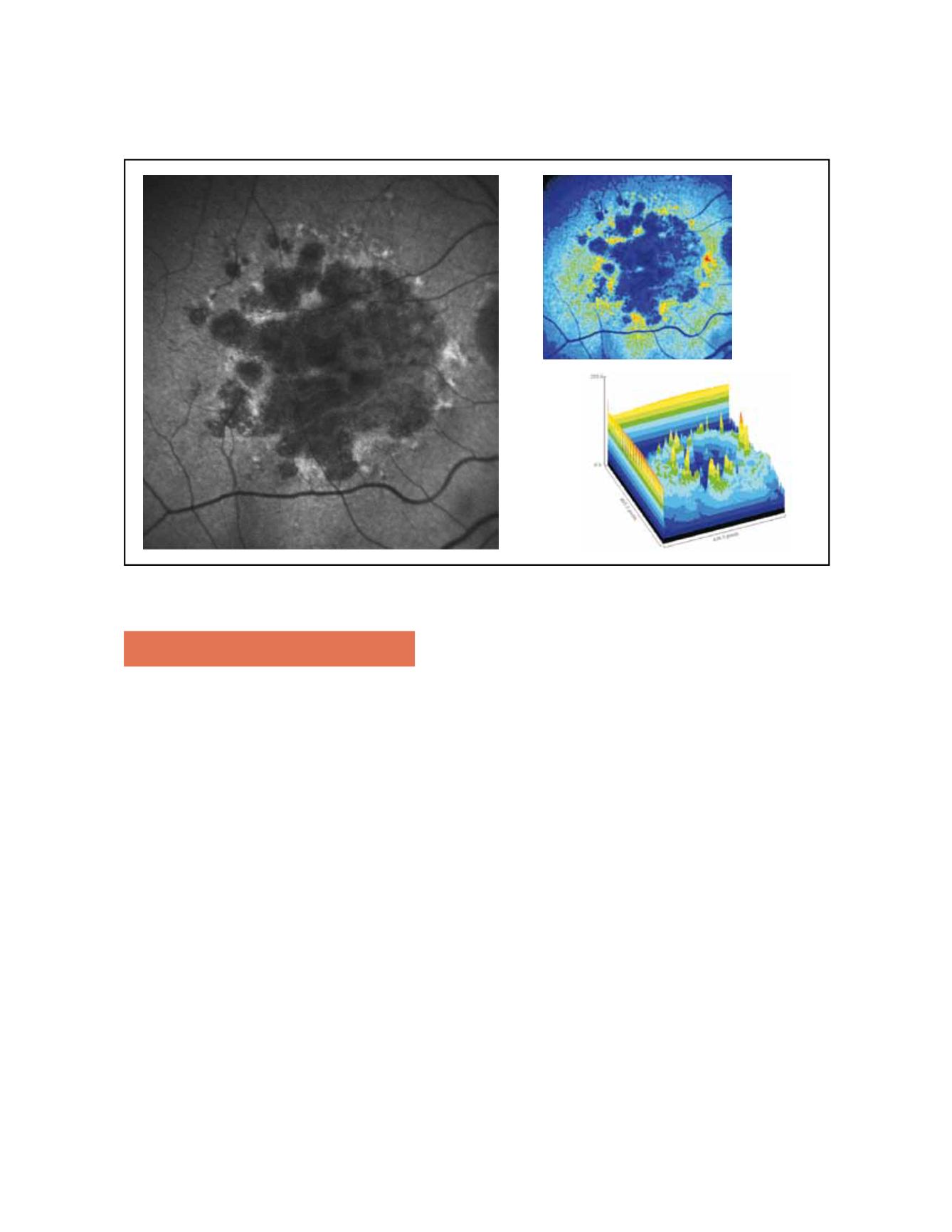
Figure 5. Surrounding areas of accumulation of lipofuscin at the junction area show increased autofluorescence. In pseudocolor 3D areas of higher
accumulation of lipofuscin are seen as yellow spikes
3. Optical coherence tomography
Recent development of high resolution, high speed spectral
domain optical coherence tomography (SDOCT) improves
the visualization of the RPE, outer and inner segment of
the photoreceptors and external limiting membrane over
previous time-domain based technology. Given the afore-
mentioned reasons, simultaneous imaging techniques that
combine SD-OCT and FAF are highly desirable for the
evaluation of the atrophic and junction areas of patients with
GA (Fig. 8). Currently there are instruments that fulfill these
criteria and allow the study of the correlation between areas
of increased or decreased autofluorescence with the morpho-
logic changes detected in the external retina by SD OCT.
SDOCT in atrophic areas of patients with GA show retinal
thinning due to atrophy of the RPE and disappearance of
the external retina, that includes inner and outer segment
of the photoreceptors and, very frequently, the external lim-
iting membrane
(18)
; in the severe forms, the outer nuclear
layer may be no longer identifiable and therefore the outer
plexiform layer may be in direct contact with Bruch´s mem-
brane. The thinned retina permits the deeper penetration of
light and a corresponding increased signal from the choroid
within the atrophic area (Fig. 9). SD OCT is also useful to
identify absence of exudative signs (intraretinal or subretinal
pockets of fluid, RPE detachments, maintenance of the con-
tinuity of Bruch´s membrane)
(19)
.
Several abnormalities have been found with SDOCT in the
junction zone
(18,20,21)
, such as disruption of external retinal
layers with different shape of band endings, disappearance
of the external limiting membrane and/or of the retina
encompassing inner segments of the photoreceptor layer to
Bruch´s membrane at the same or at a different transverse
planes, small elevations of RPE thought to represent sub-
RPE deposits or increased distance between inner and outer
photoreceptors segment and RPE, presumably due to debris
between these layers. Smooth margins with no structural
changes from normal to abnormal retina exist in the junc-
tion zone when there is no FAF abnormality, which under-
scores the significance of abnormal FAF
(21)
(Fig. 9).
In summary, precise quantification of theGA and its progres-
sion by fundus autofluorescence imaging and the detailed
morphologic study of the external retina by the high resolu-
tion SD OCT allow to show the relative slow progressing
abnormalities of the outer retinal layers in dry AMD. This
may prove essential for prognostic and interventional strate-
gies, in order to detect potential benefit by slowing down the
degenerative process or perhaps detect signs of RPE and/or
photoreceptors rescue.


