120
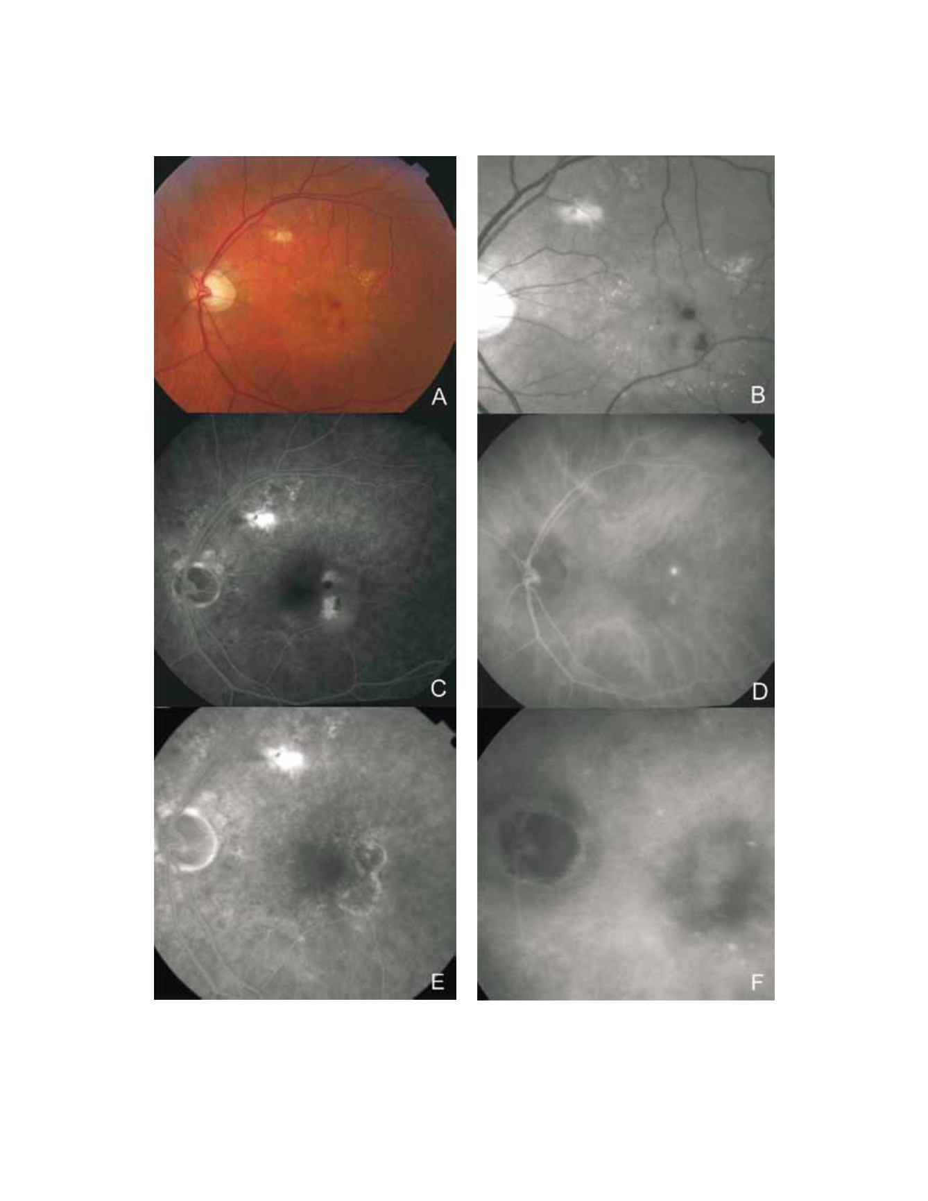
Figure 3 - RAP lesion. Fundus colour photography (A) with intra-retinal haemorrhages, hard exudates and neurosensory detachment.
Red-free (B) with two juxtafoveal and extrafoveal small haemorrhages. Fluorescein angiography (C) shows two juxtafoveal hyperfluo-
rescent spots, neurosensory detachment and pigment epithelium detachment. Late ICG reveals two juxtafoveal hyperfluorescent hot
spots. E and F: Fluorescein angiography and ICG after laser photocoagulation with resolution of exudation.


