121
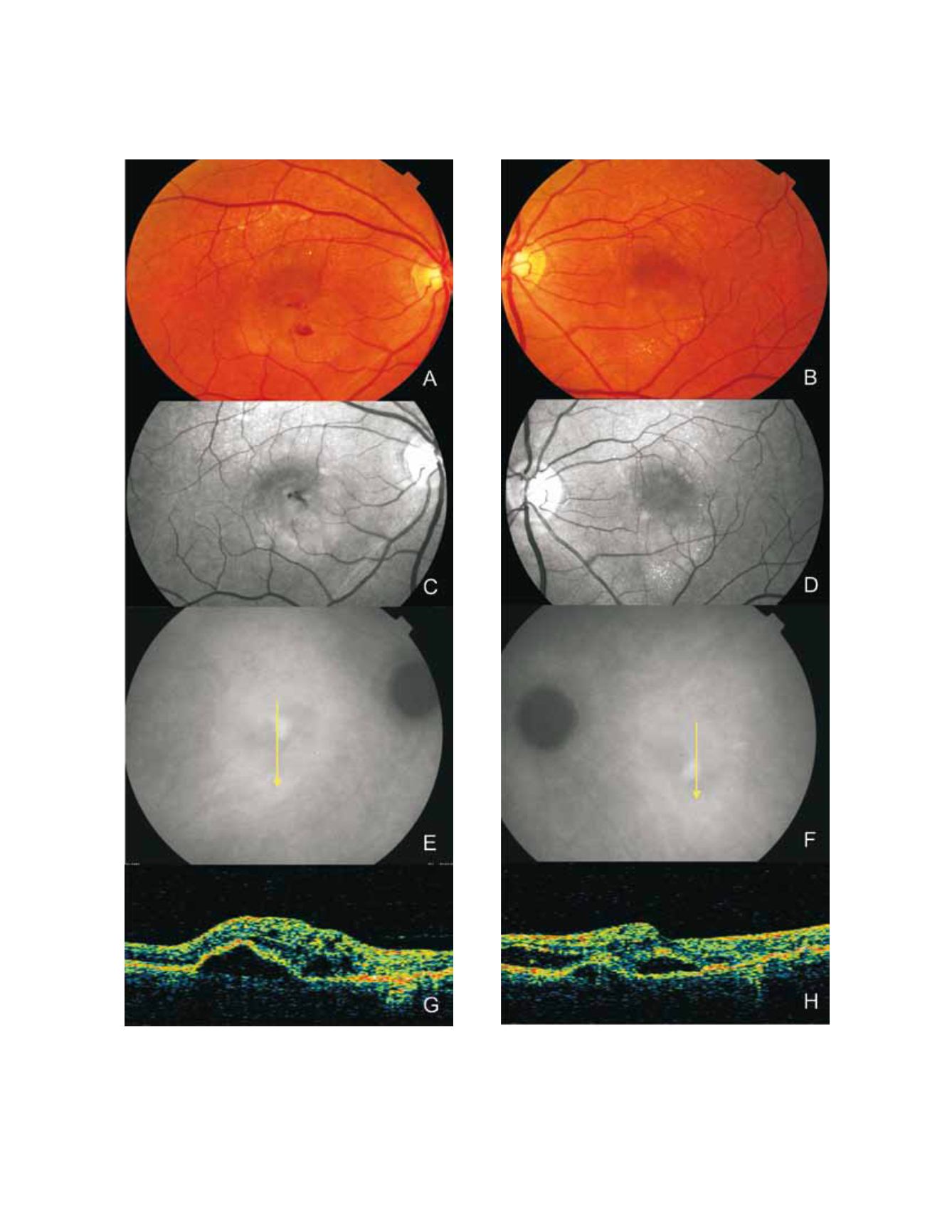
Figure 4 - Bilateral RAP lesion: Fundus colour photography and red-free images (A,B,C,D) clearly show retinal oedema, small retinal haemorrhages
and lipidic exudation. E and F: late ICG both eyes with a hot spot and subfoveal hypofluorescence (serous PED). Stratus OCT reveals, in both eyes
(G and H), serous PED, neurosensory detachment and intra-retinal fluid.
Neovascular Phenotypes: RAP (Retinal angiomatous proliferation)


