114
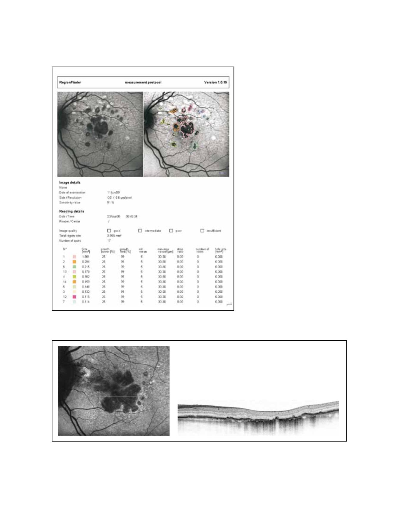
Figure 7. Semiautomated software allows quantification of total area of GA and measurement of its progression in time (Spectralis Heidelberg Retinal
Angiograph/OCT; Heidelberg Engineering, Heidelberg, Germany )
Figure 8 - High resolution optical coherence tomography correlated with the autofluorescence image.


