109
Fundus autofluorescence patterns and
optical coherence tomography in
geographic atrophy secondary to AMD
10
Geographic atrophy (GA) and choroidal neovasculariza-
tion (CNV) represent the advanced forms of age-related
macular degeneration (AMD). GA is defined as a well
circumscribed area of atrophy of the retinal pigment epi-
thelium (RPE) where the large choroidal vessels can be
seen by ophthalmoscopy and show thinning or absence
of the RPE, closure of the choriocapillaris and degenera-
tion of the overlying photoreceptors
(1,2)
.
Visual loss in GA is due to areas of atrophy of the RPE
larger than 175 µm and subsequent loss of tissue in the
outer retina (photoreceptors) and choriocapillaris; these
areas tend to coalesce progressively and may not affect the
fovea until late in the course of the disease (the so-called
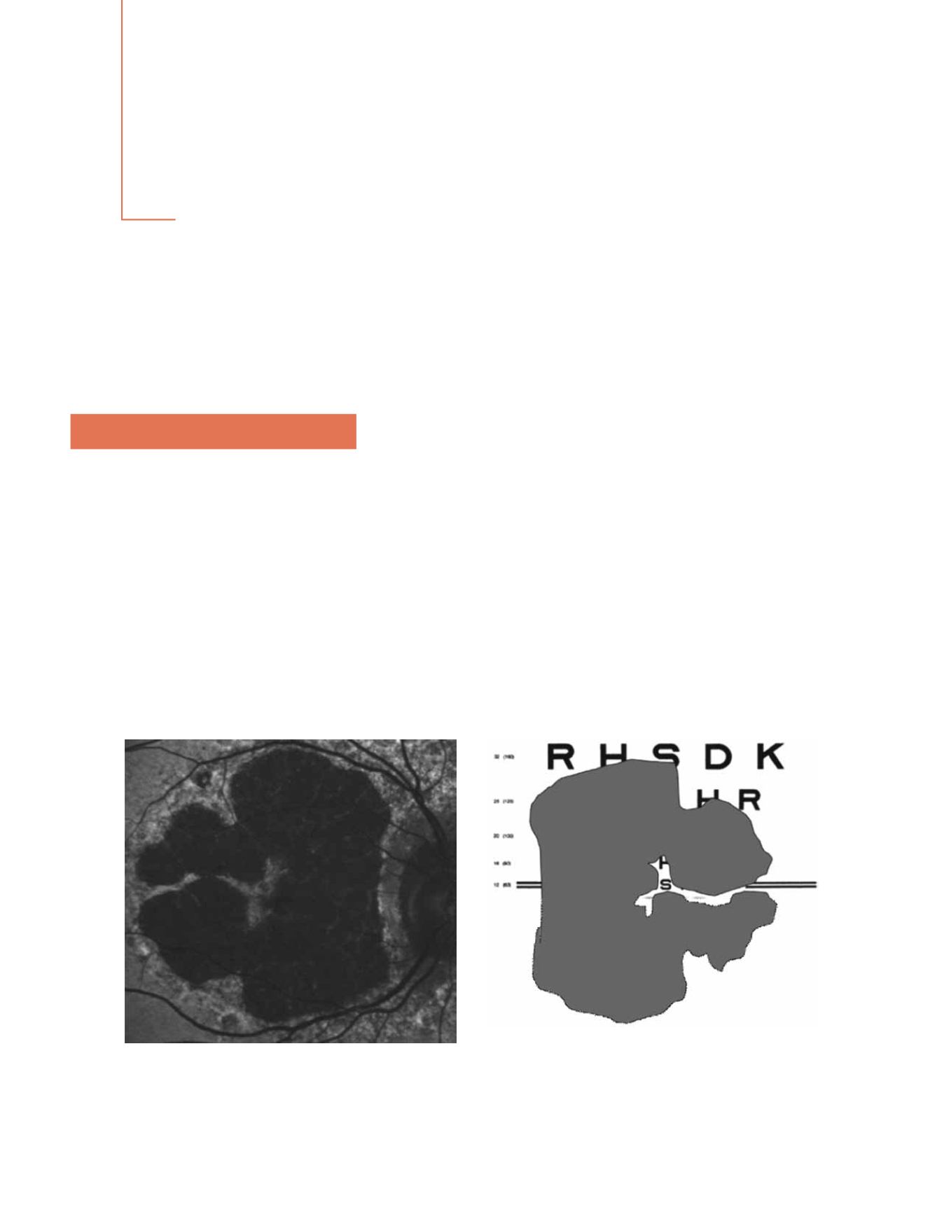
“foveal sparing”), when visual acuity (VA) finally ensues.
Due to the large paracentral areas of atrophy but preser-
vation of the fovea visual function is often very poor in
spite of an apparently good VA. Patients with a VA of
20/20 may be functionally blind (Fig. 1). Relatives of the
patients, and even ophthalmologist, have often confused
VA for visual function, thus, frequently these patients
have felt poorly understood
(1)
. GA is responsible for one-
third of the cases of end stage disease
(3)
and accounts for
20% of cases of severe visual loss due to the disorder
(4)
.
At the age of 85 years or older incidence of GA is four
times the one of CNV. GA is not a benign disease; the
atrophy of the external layers may progress at a speed of
1.5-2.6 mm² per year (Fig. 2)
(5)
.
1. Introduction
Authors:
Jordi Monés, MD
1
Marc Biarnés, OD, MPH
1
1
Institut de la Màcula i de la Retina, Centro Médico Teknon, Barcelona. Spain
Figure 1: Foveal sparing may allow a 20/20 visual acuity in spite of very severe visual function impairment


