115
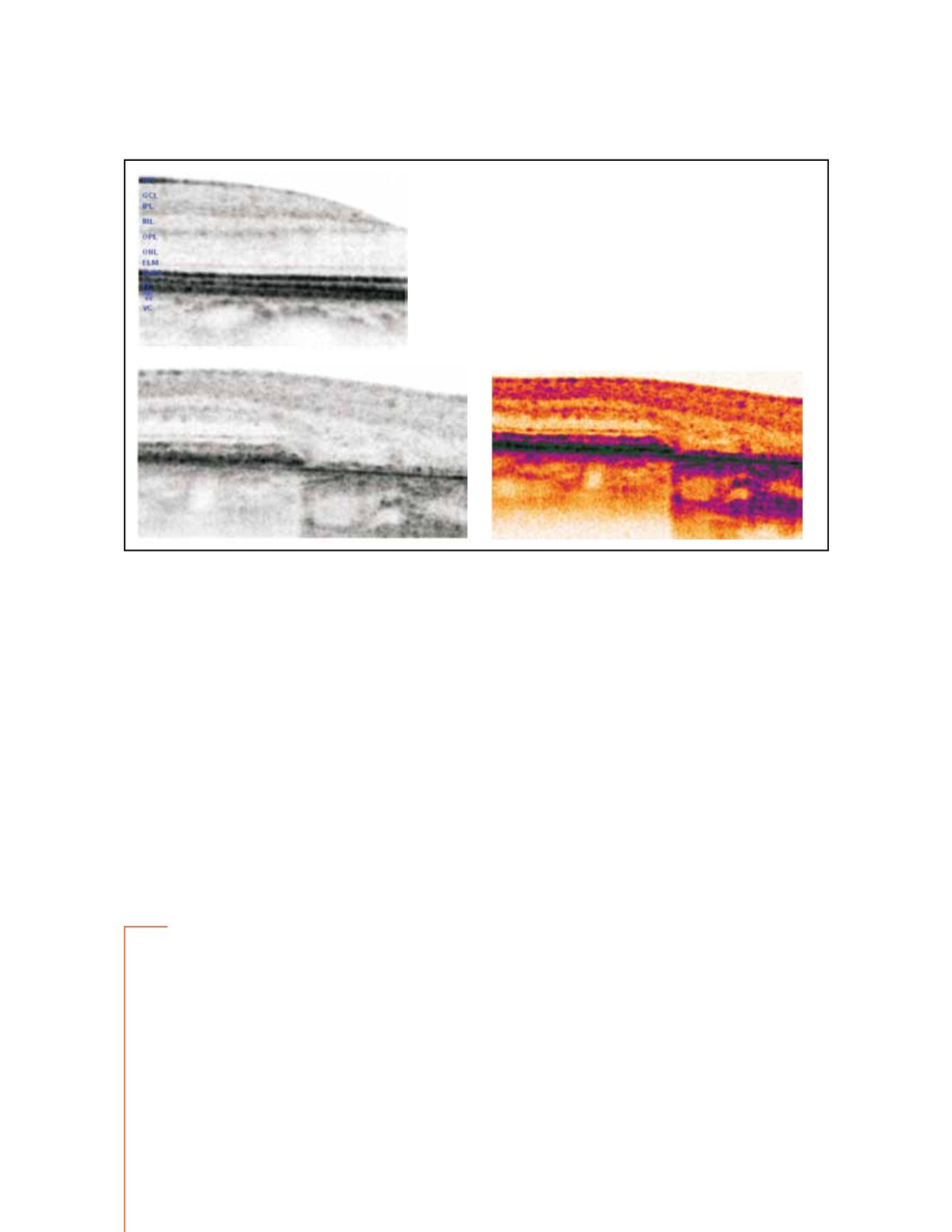
Figure 9 - SD OCT in normal retina (up left ). In the lower images, SD OCT of the junction area between preserved retina and the geographic
atrophy. Atrophic areas show retinal thinning due to atrophy of the RPE and disappearance of the external retina that includes inner and outer
segment of the photoreceptors and, very frequently, the external limiting membrane
(16)
; The outer nuclear layer may be no longer identifiable and
therefore the outer plexiform layer may be in direct contact with Bruch´s membrane. The thinned retina permits the deeper penetration of light and
a corresponding increased signal from the choroid within the atrophic area.
Correspondence concerning this article can be sent directly to the authors through the emails:
Fundus autofluorescence patterns and optical coherence tomography in geographic atrophy secondary to AMD


