103
Geographic Atrophy
as a clinical marker for changes in Bruch’s membrane,
and as a risk factor for development of geographic
atrophy
(24)
. Despite these aspects in GA, fluorescein
angiography may be indicated only in atypical cases,
in order to allow the correct diagnosis
(38)
.
5.3 Optical coherence tomography (OCT)
OCT scan shows thinning of hyperreflective external
band, corresponding to attenuation of RPE/Bruch’s
complex, and deeper hyperreflectivity because of loss
of outer layers including photoreceptors (Fig. 4)
(39,40)
.
In high resolution OCT the atrophic area shows
hyperreflective clumps at different levels, segmented
plaques of the outer band and elevations with vari-
able reflectivity
(41)
.
In the perilesional area there are elevations of the
outer retinal layers, as well as thickening of outer
hyperreflective band. At the junction area the outer
band shows different degrees of loss
(41)
.
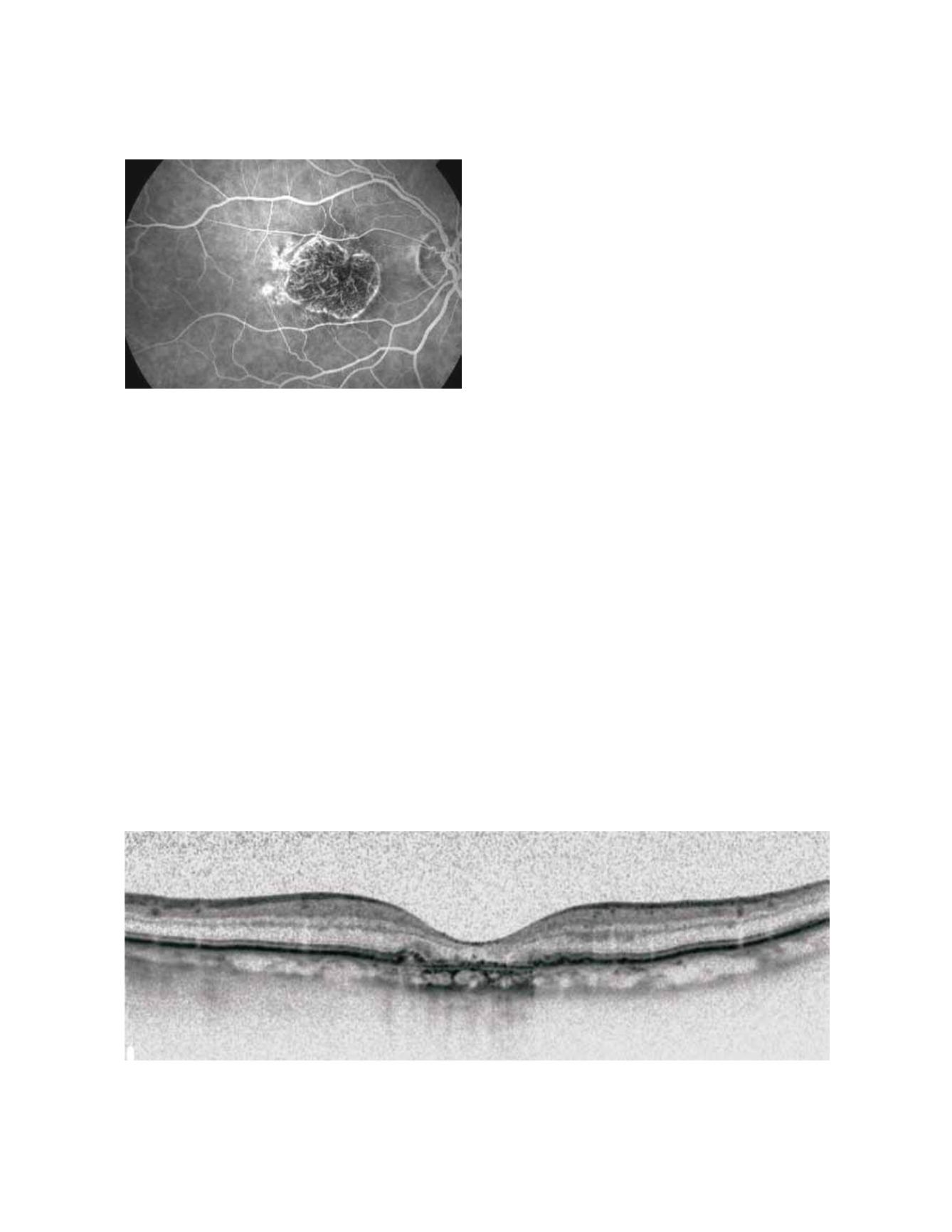
Figure 3.
Fluorescein angiography showing a sharply delineated window defect.
Figure 4. Spectralis OCT: Thinning of hyperreflective external band (because of attenuation of RPE/Bruch’s complex) and deeper
hyperreflectivity.
5.4 Fundus autofluorescence (FAF)
Fundus spectrophotometric studies
in vivo
by Delori
and co-workers, have shown that FAF represents
an accumulation of lipofuscin in the lysossomes of
RPE cells, mainly derived from photoreceptors outer
segments degradation. The compound is found as
micrometer-sized spherical particles and is character-
ized by yellow autofluorescence when exposed to blue
light
(42,43,44)
.
It has been shown with confocal scanning laser oph-
thalmoscopy (cSLO) that FAF response is very low
or extinguished in areas of atrophy. The lack of RPE
cells or its low number and therefore of lipofuscin,
(the dominant fluorophore) explain this reduction
(45)
.
Increased FAF precedes development of GA
(46,47)
.
FAF is increased in junctional zone around areas of
atrophy, and intensity seems to correlate with exten-
sion of the atrophic area, and also with reduction of
retinal sensitivity detected by fundus perimetry
(48,49)
.


