95
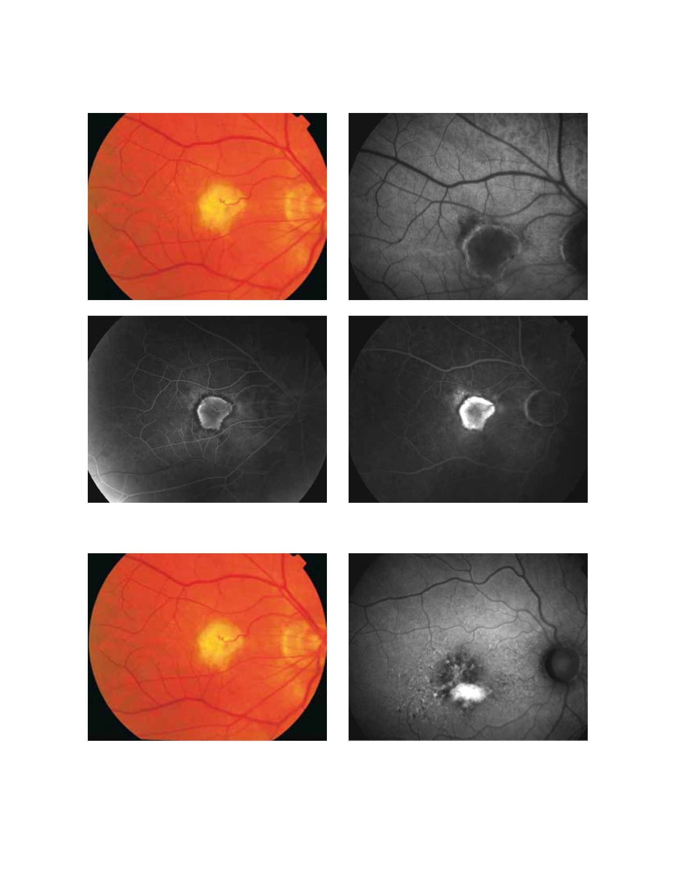
Figure 17 - Choroidal neovascularization with chorioretinal anastomoses. (A) Colour fundus and (B) fundus autofluorescence photographs. (C)
Early frame fluorescein angiography. (D) Late frame fluorescein angiography.
Figure 18 - Retinal pigment epithelial detachment. (A) Colour fundus and (B) fundus autofluorescence photographs.
Fundus autofluorescence in age-related macular degeneration
A
B
C
D
A
B


