91
Fundus autofluorescence in age-related macular degeneration
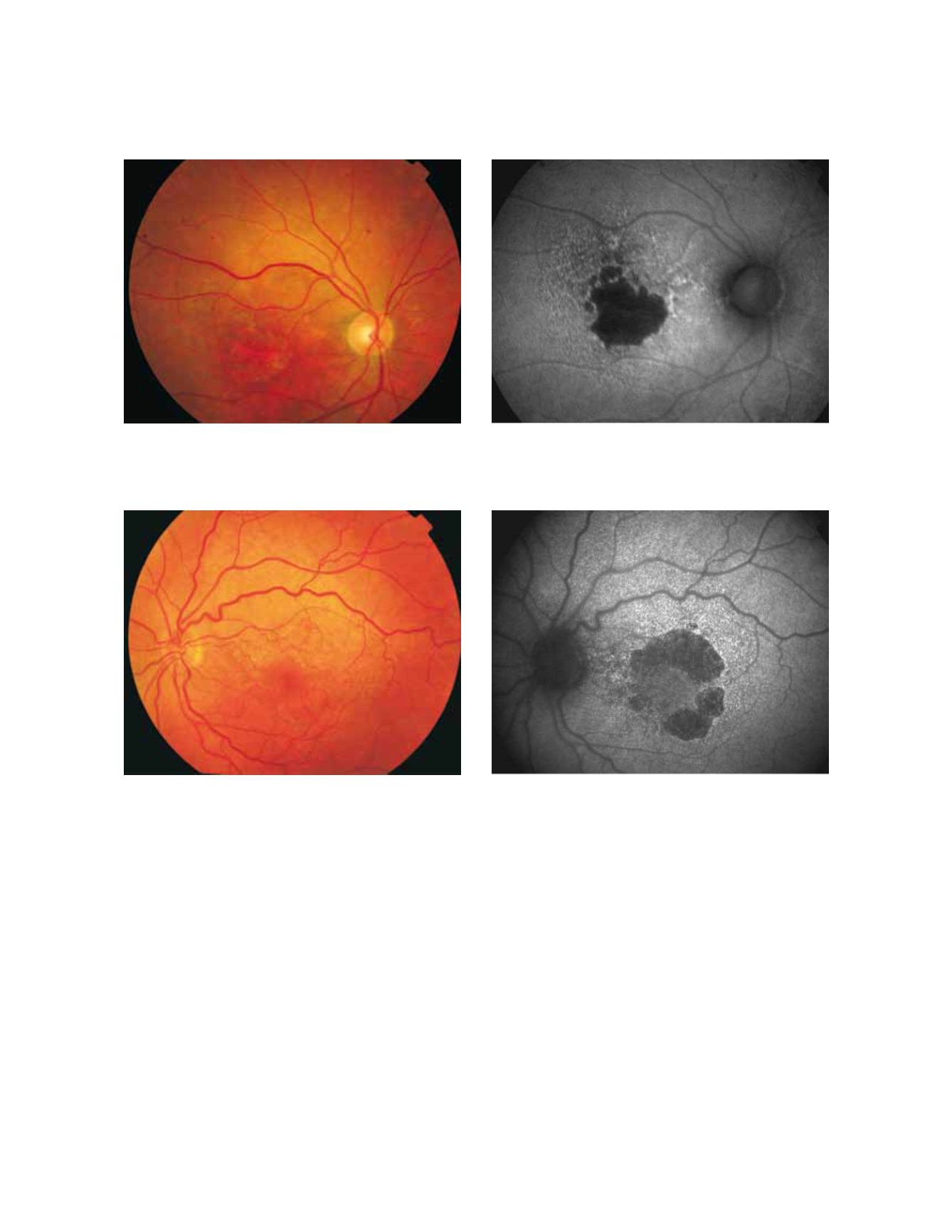
Figure 12 - Geographic atrophy (GA) secondary to AMD with increased autofluorescence in the junction. New areas of GA and the extension of
pre-existing areas are characterised by an excessive accumulation of LF, and therefore an increased FAF signal. (A) Colour fundus and (B) fundus
autofluorescence photographs.
Figure 13 - Geographic atrophy secondary to AMD with a banded pattern of increased autofluorescence in the junction. (A) Colour fundus and
(B) fundus autofluorescence photographs.
A
B
with a fine branching pattern of increased FAF.
-Fine granular pattern,
is defined by a large area of
increased FAF with a granular appearance surrounding
the GA area and a clear border between the granular
increased FAF and the surrounding normal background
FAF.
-Fine granular with peripheral
punctate spots pattern
is characterised by diffuse FAF changes surrounding the
atrophic area with elongated small lesions and increased
FAF.
Refined phenotypes help to identify the prognosis and
seem to be a prerequisite to determine specific genetic
factors in a complex, multifactorial disease such as AMD.
A recent analysis of the follow-up of junction FAF pat
terns in GA and the rate of progression of atrophic lesions
revealed that variation in GA growth rates are dependent
on the specific phenotype of FAF at baseline
(49)
. Atrophy
enlargement was slowest in eyes with normal FAF pat-
tern (median, 0.38 mm2/year), followed by focal FAF
pattern (median, 0.81 mm2/year), diffuse FAF pattern
(median, 1.77 mm2/year), and banded FAF pattern
(median, 1.81 mm2/year). The rate of progression of
GA in eyes with patchy FAF pattern were not included
in this analysis because of their low frequency, insuf
ficient for statistical analysis.The rate of progression of
“banded” and “diffuse” FAF patterns were significantly
A
B


