84
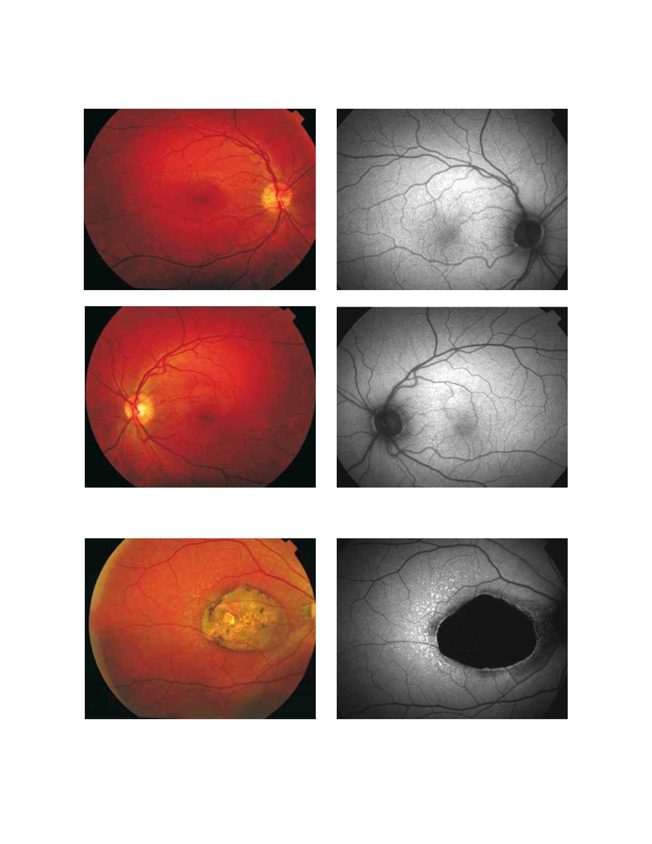
Figure 2 - Atrophic area of RPE. These areas typically appear as dark patches in FAF images and can be clearly delineated. (A) Colour fundus and
(B) fundus autofluorescence photographs.
A
A
C
B
B
D
Figure 1: Colour fundus and fundus autofluorescence from a normal subject. (A) Right eye colour fundus photograph and (B) fundus autofluores-
cence. (C) Left eye colour fundus photograph and (D) fundus autofluorescence.


