90
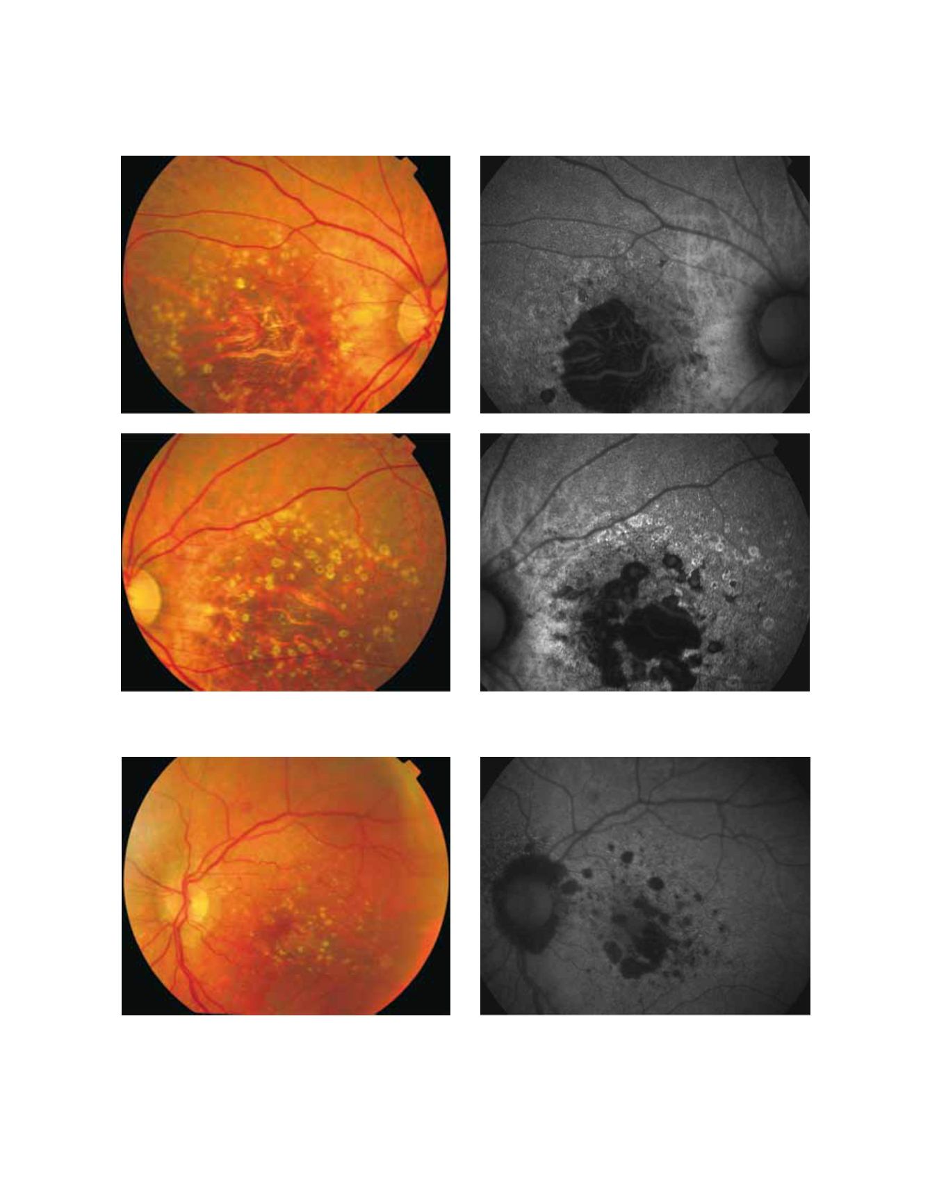
Figure 10 - Geographic atrophy secondary to AMD. The loss of RPE and LF causes a characteristic absence of FAF signal (a dark area in FAF images).
(A) Right eye colour fundus photograph and (B) fundus autofluorescence. (C) Left eye colour fundus photograph and (D) fundus autofluorescence.
A
B
C
D
Figure 11 - Geographic atrophy secondary to AMD. The high contrast between atrophic and non-atrophic retina enables the delineation of the
atrophic area more precisely than can be performed from conventional fundus photographs. (A) Colour fundus and (B) fundus autofluorescence
photographs.
A
B


