86
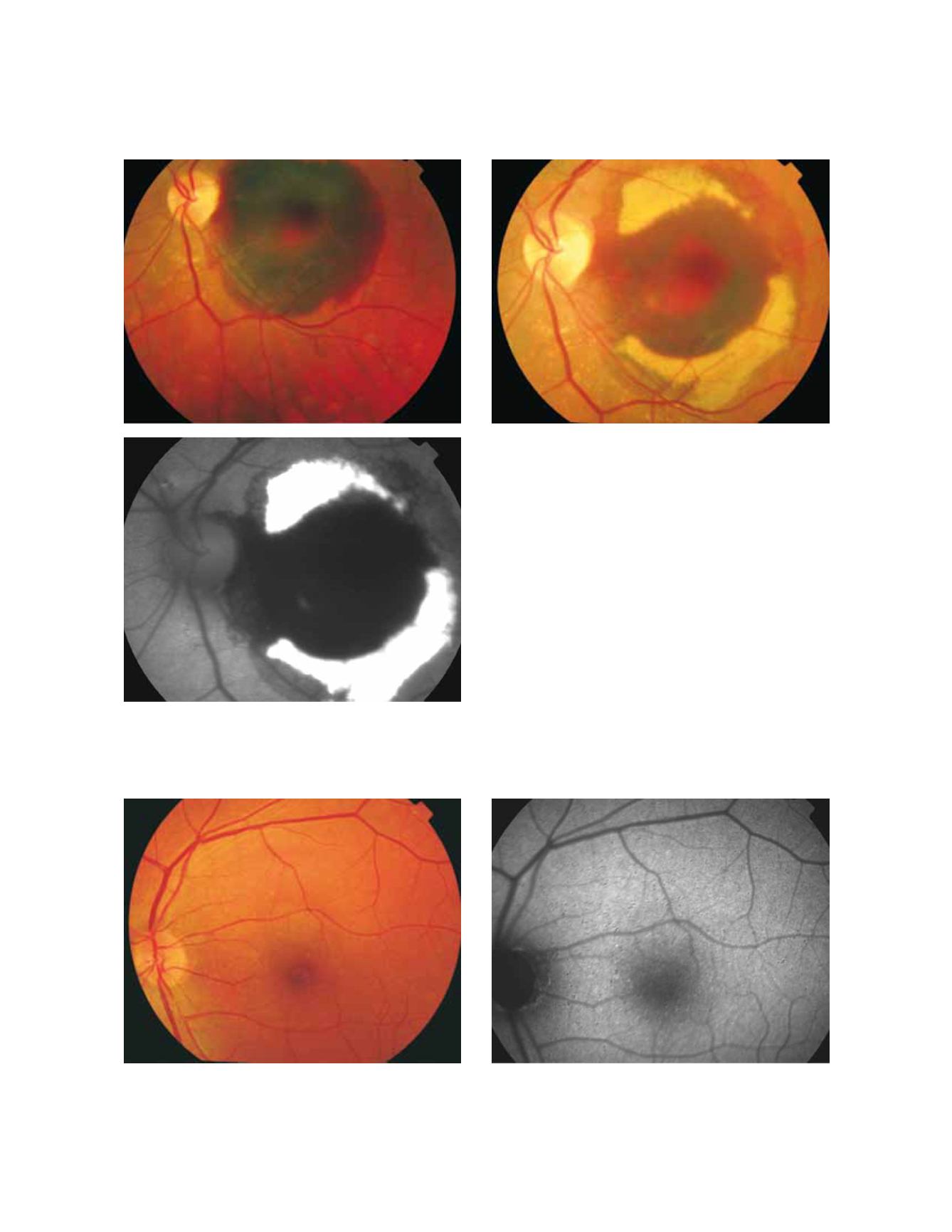
Figure 5 - Small drusen from a patient with early AMD in the left eye. (A) Colour fundus and (B) fundus autofluorescence photographs.
A
B
C
Figure 4 - Haemorrhage in the left eye of a patient with choroidal neovascularization secondary to AMD. Fluorophores eventually appear in haemor-
rhagic areas which are observed as yellowish areas in the fundus and as an increased FAF signal. (A) Colour fundus photograph of the fresh haemor-
rhage. (B) Colour fundus photograph from the same eye one month later. (C) Fundus autofluorescence of the haemorrhage.
A
B


