251
inner and outer segments of the photoreceptors and
higher VA in patients treated with ranibizumab.
63-64
It appears that parallel to the development of new
devices, structures assume special importance, such
as the junction of the inner and outer segments of
the photoreceptors, which until now was only identi-
fied, as shown by studies with prototypes with high-
definition axial resolution of 3.5 microns as we will
discuss later in this chapter.
65-66
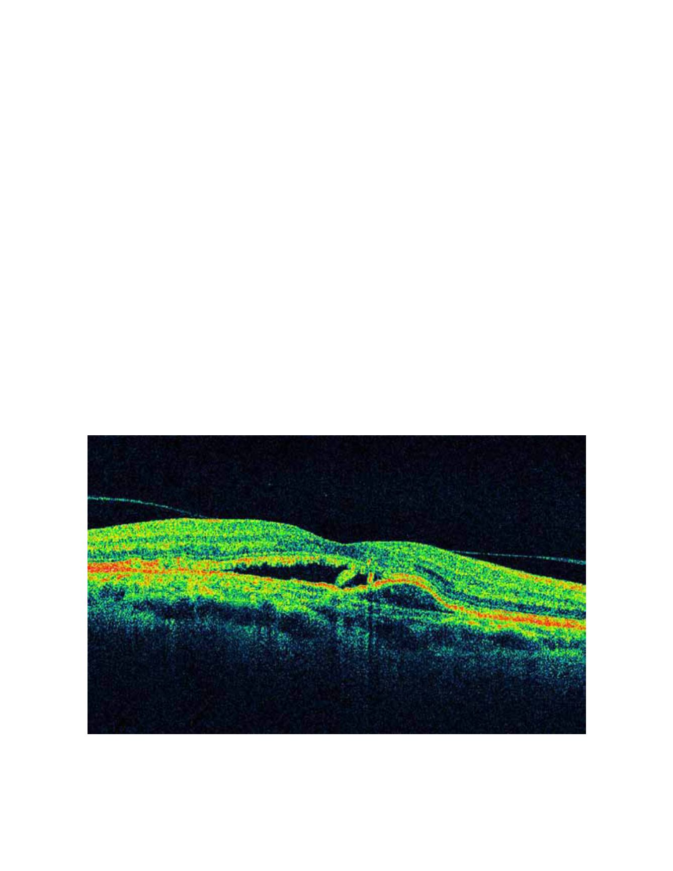
Lee and colleagues
reported that the presence of posterior vitreomacu-
lar adhesion on OCT was associated with CNV in a
large series of patients with AMD (n = 251). Those
authors suggested that this finding is a possible risk
factor for subretinal membrane development because
of chronic vitreomacular traction on the retina,
opening the door to a possible surgical approach in
patients not responding or resistant to drug treatment
(Figure 21).
67-68
previous treatments.
60
Keane and colleagues also
found that the main factor associated with decreased
VA was the volume of subretinal tissue, and in fewer
cases, thickening of the neurosensory retina, without
a significant association with the total volume of sub-
retinal fluid and RPE detachment and VA.
61
However,
these parameters did not justify the variability found
in the VA for similar values of subretinal tissue vol-
ume or thickening of the neurosensory retina. The
authors pointed to the complex pathophysiology of
the neovascular membranes and the limitations of
the TD-OCT used to explain the results. Sayanagi
and colleagues reported that SD-OCT is a superior
generation of TD-OCT than its predecessors for
assessing the activity of the neovascular membranes
and changes in AMD after ranibizumab treatment,
and Kiss and colleagues pointed to the RPE status
of neovascular membranes as the main predictor of
VA in patients treated with ranibizumab in addition
to the conventional parameters such as central reti-
nal thickness.
37-62
Several studies also reported a sig-
nificant correlation between a hyperreflective band
indicating the integrity of the junction between the
Figure 21.
The chronic retinal vitreomacular traction has been considered as a possible risk factor for development of subretinal
membranes. In the image, a subretinal membrane associated with a vitreomacular traction in the presence of an RPE detachment
and subretinal fluid may be appreciated.
OPTICAL COHERENCE TOMOGRAPHY IN AGE-RELATED MACULAR DEGENERATION


