245
Figure 15.
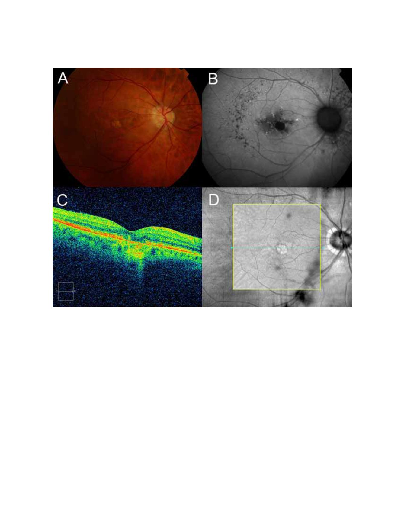
(A) The retinography shows a juxtafoveal atrophic plaque. (B) The autofluorescence image defines the atrophic plaque
as an area of complete absence of fluorescence. (C) The OCT shows an increased choroidal reflectance due to the window effect
produced by the overlying atrophic plaque. (D) Infrared image captured by the OCT that shows the tomographic section of figure
C.
OPTICAL COHERENCE TOMOGRAPHY IN AGE-RELATED MACULAR DEGENERATION


