241
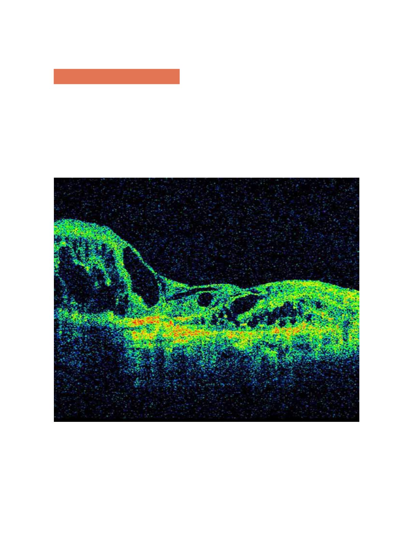
Figure 12.
OCT shows a hyperreflective fibrotic nodular area that corresponds to the fibrotic area and easily detects diffuse or
cystoid edema. In the wet form, OCT shows diffuse or cystoid edema involving the RPE detachment.
10. Disciform scars
Disciform scars form the stage of exudative AMD in
which the retinal tissue is replaced by scar tissue (usu-
ally vascular). Disciform scars develop with regres-
sion of subretinal hemorrhages and retinal edema and
hyperplasic elements of the RPE. Disciform scars are
usually dry and show marked destruction of all retinal
layers. It is less common to find diffuse exudation with
elevation of the entire sensory retina at the posterior
pole (Figure 12).
OPTICAL COHERENCE TOMOGRAPHY IN AGE-RELATED MACULAR DEGENERATION


