232
3. Atrophic Macular Degeneration
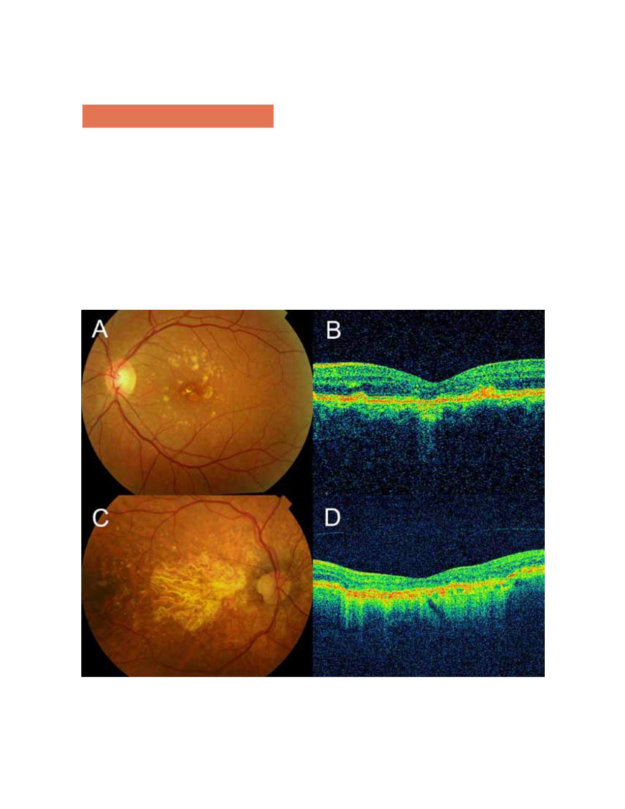
In the macular region, progressive atrophy of the RPE,
the outer retinal layers, the choriocapillaris, and dense
clusters of drusen can be seen.
OCT can detect decreases in retinal thickness and
increases in the reflectivity of the RPE detachment,
which results from the decreased ability of the atrophic
retinal tissue to attenuate light. The reduced retinal
thickness and volume can be determined by the retinal
maps that display the areas of greatest atrophy, iden-
tify the extent of the atrophy, and monitor progression.
Geographic atrophy (GA) represents the final stage of
dry AMD.
6,8,10,16,17
Figure 3.
In a case of retinal atrophy, the OCT shows a highly reflective choroidal signal due to retinal thinning and RPE hypop-
igmentation, which allows greater beam penetration into the choroid and greater reflectivity. The retinal map allows quantification
of the decreased retinal thickness.


