233
4. Exudative Macular Degeneration
OCT, a fundamental tool in the diagnosis and man-
agement of patients with choroidal neovasculariza-
tion (CNV), allows identification of active neovascu-
lar membranes and determination of the extent of the
membranes in many cases. The technology is useful for
assessing subfoveal involvement and also helps diagnose
hidden choroidal neovascular membranes, for which
fluorescein angiography (FA) shows confusing patterns.
OCT also can monitor treatment responses by provid-
ing information about the need for retreatment.
3,8,15,18-23
Well-defined classic CNV appears on OCT as hyper-
reflective areas in contact with or in front of the RPE;
the pathology may be dome-shaped or appear as a thin
formation (fusiform or nodular) (Figure 4). Retinal
edema is always present to some extent in front of the
active membrane; if the retina is thinner than normal,
new vessels may be latent. CNV is less evident a few
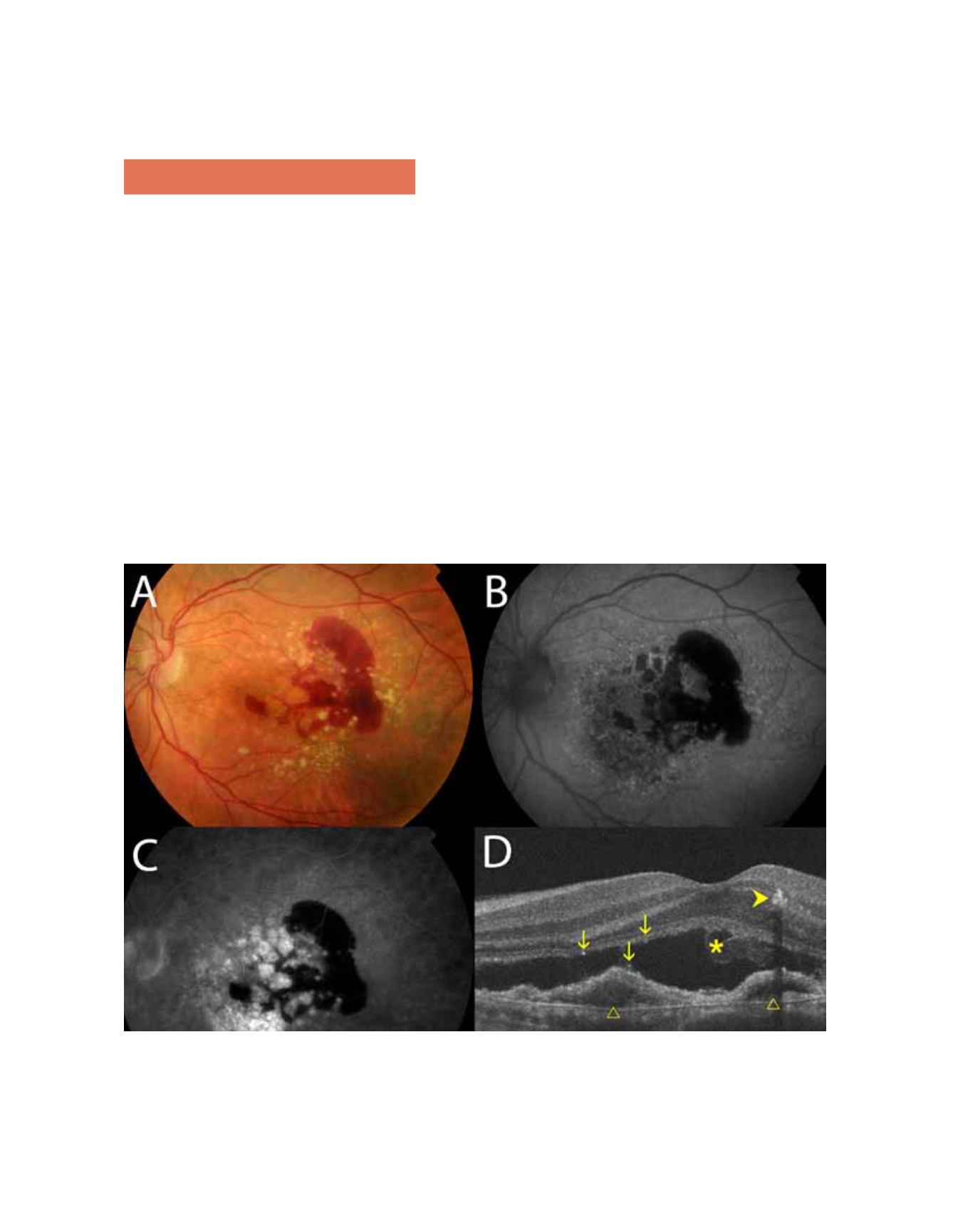
Figure 4.
OCT visualizes the components of the neovascular membrane, a retinal pigment epithelial detachment (PED), detach-
ment of the neuroepithelium (NED), intraretinal fluid, and subretinal hemorrhage.
weeks after onset, and only interruption, breakdown,
and pronounced thickening of the RPE can be seen.
OPTICAL COHERENCE TOMOGRAPHY IN AGE-RELATED MACULAR DEGENERATION


