240
Although the initial stages are only visible by fun-
dus examination and angiographic examinations
due to the small size of the initial injury, OCT is
extremely useful for identifying RAP and its associated
manifestations.
6-8,19,27-29
The initial signs that correspond to stage 1 (intraretinal
vascularization) consist of a focal area, usually extrafo-
veal, with increased retinal reflectivity that are not associ-
ated with epiretinal, intraretinal, or subretinal changes or
changes in the retinal thickness. Progression of RAP on
OCT is characterized by the presence of intraretinal or
subretinal fluid, the former characterized by well-defined
confluent hypo-reflective spaces and the latter (neuro-
sensory detachment) characterized by a well-defined
hypo-reflective space between the neurosensory foveal
retina and other highly reflective bands corresponding
to the RPE. When RAP reaches the subretinal space and
merges with the RPE, a serous detachment of the RPE
usually develops (stage 2 or CNV). In well-developed
cases, there may be retinal choroidal anastomoses (stage
3 or CNV) and it is impossible to clearly differentiate
stage 2 from stage 3 on OCT.
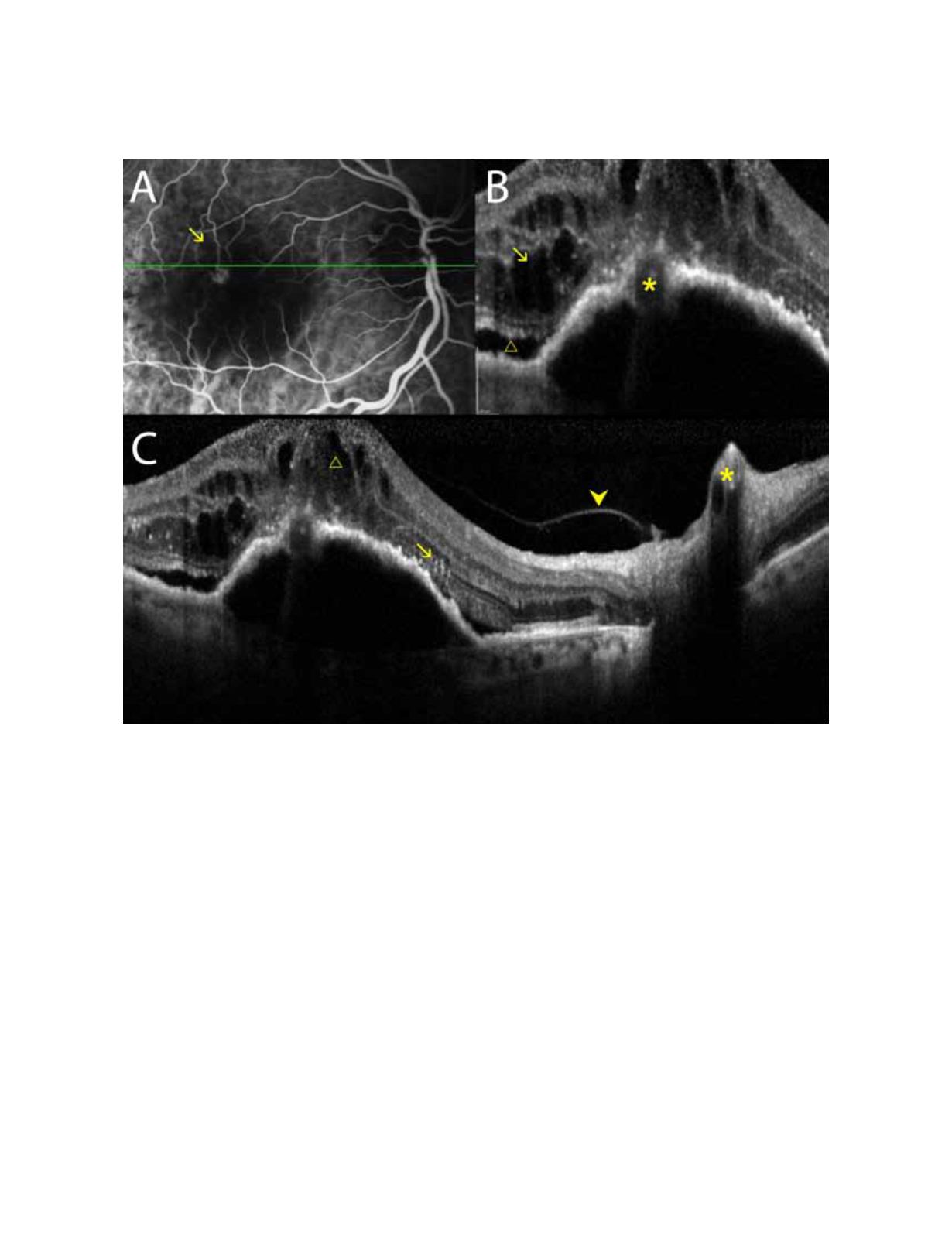
Figure 11.
A and B, OCT shows RAP associated with edema, intraretinal cysts, and subretinal fluid. C, OCT shows the RPE and
the retinal neurosensory layer detachments.


