244
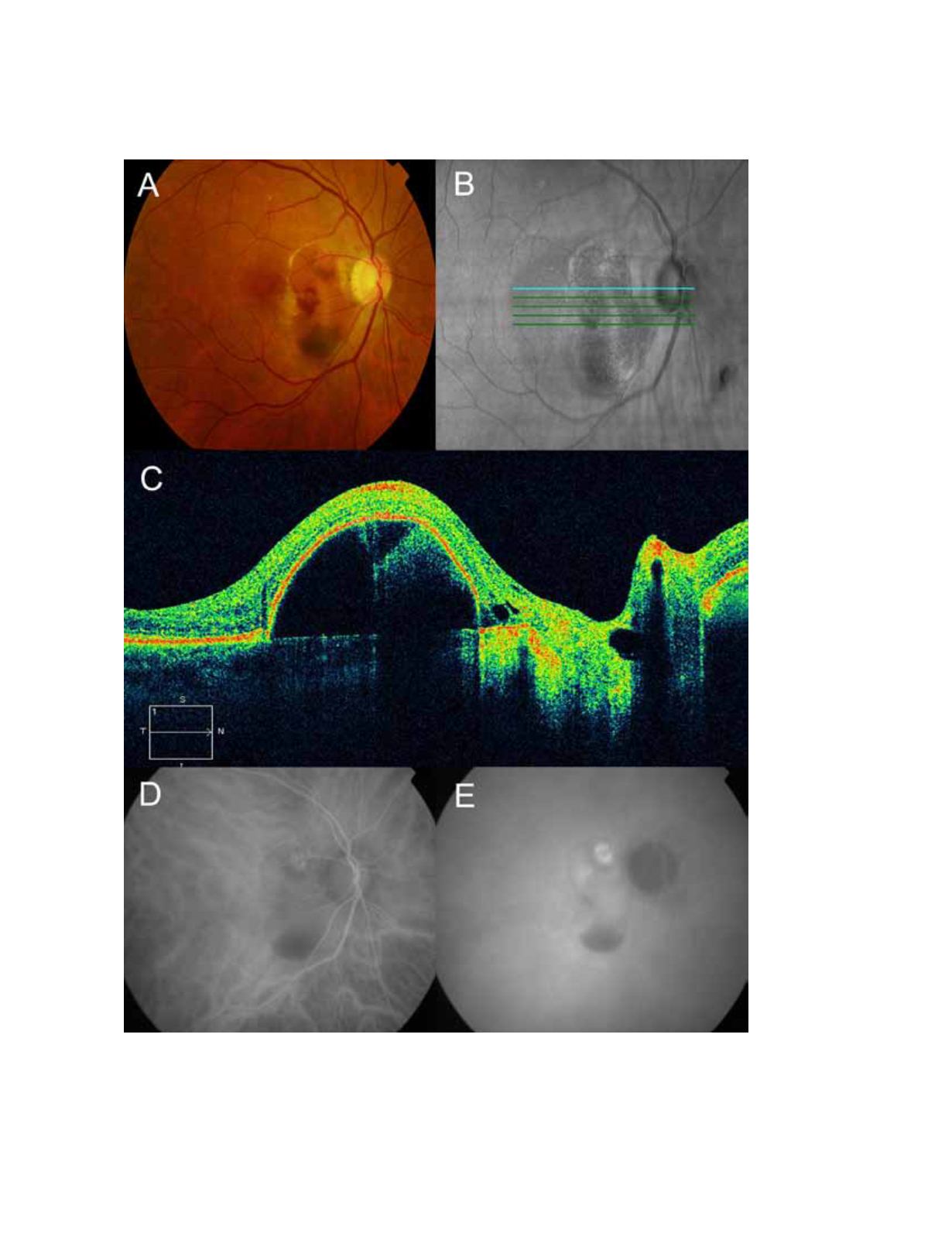
Figure 14.
The retinography corresponds to a polypoidal choroidopathy (A) where you can observe the presence of intraretinal
haemorrhages. In the red-free image (B) you can observe the tomographic cut line. The OCT shows a marked elevation of the
cup-shaped RPE (C). By indocyanine green angiography you can observe a hot spot that confirms the diagnosis (DE).


