246
14. Clinical and Therapeutic Implications of
OCT in AMD
OCT is now a routine examination for patients with
AMD. Although some studies have advocated a role for
OCT in the dry forms of the disease, its use is greater in
patients with the neovascular forms, and it is now a main
method of study in these patients. From the first studies
of PDT, the different patterns of response to treatment
were assessed with OCT, and the development of antian-
giogenic therapies made OCT a fundamental tool in
routine patient management. In recent years, OCT has
become increasingly important, to the extent that recent
studies have based the need for retreatment on the find-
ings in OCT images.
15. Dry AMD
The better definition achieved with the OCT devices
that include SD technology allows more detailed study
of the outer retinal layers and RPE, which are impor-
tant structures in the development of dry AMD. Thus,
new SD-OCT instruments can accurately distinguish
the presence and size of drusen and RPE changes, mak-
ing it possible to differentiate the different retinal lay-
ers to identify changes at that level.
48
Thus, in a group
of patients with dry AMD, Schuman and colleagues
49
found localized thinning of the photoreceptor layer
immediately above the drusen compared to healthy
controls, suggesting a degenerative process with cell loss
to explain the decreased visual function in this group
(Figure 16).
B
earelly and colleagues
17
studied the thinning at the
edges of the plaques of GA to establish a gradient in the
thickness of the photoreceptors layer from the healthy
retina to the atrophic plaque. Although they had a small
sample size in their study (n = 17), they concluded that
SD-OCT allows quantitative measurement of disease
progression and postponed for future study the applica-
tion of the technique. Moreover, to study progression of
the dry forms, other studies have been designed to cor-
relate the findings with the SD-OCT scans with other
techniques such as the autofluorescence mentioned pre-
viously. To this end, Stopa and colleagues
47
analyzed a
series of patients with dry AMD and correlated SD-OCT
images of areas of GA, and isolated hard drusen and soft
coalescing drusen with retinographies and images with
autofluorescence. Thus, in addition to finding differ-
ent patterns of reflectivity for each type of drusen, they
observed that certain patterns of hyperreflectivity in
some drusen and the overlying retina corresponded to
increased or decreased autofluorescence at these points.
This established a certain morphology-function relation-
ship with both scans (Figure 17).
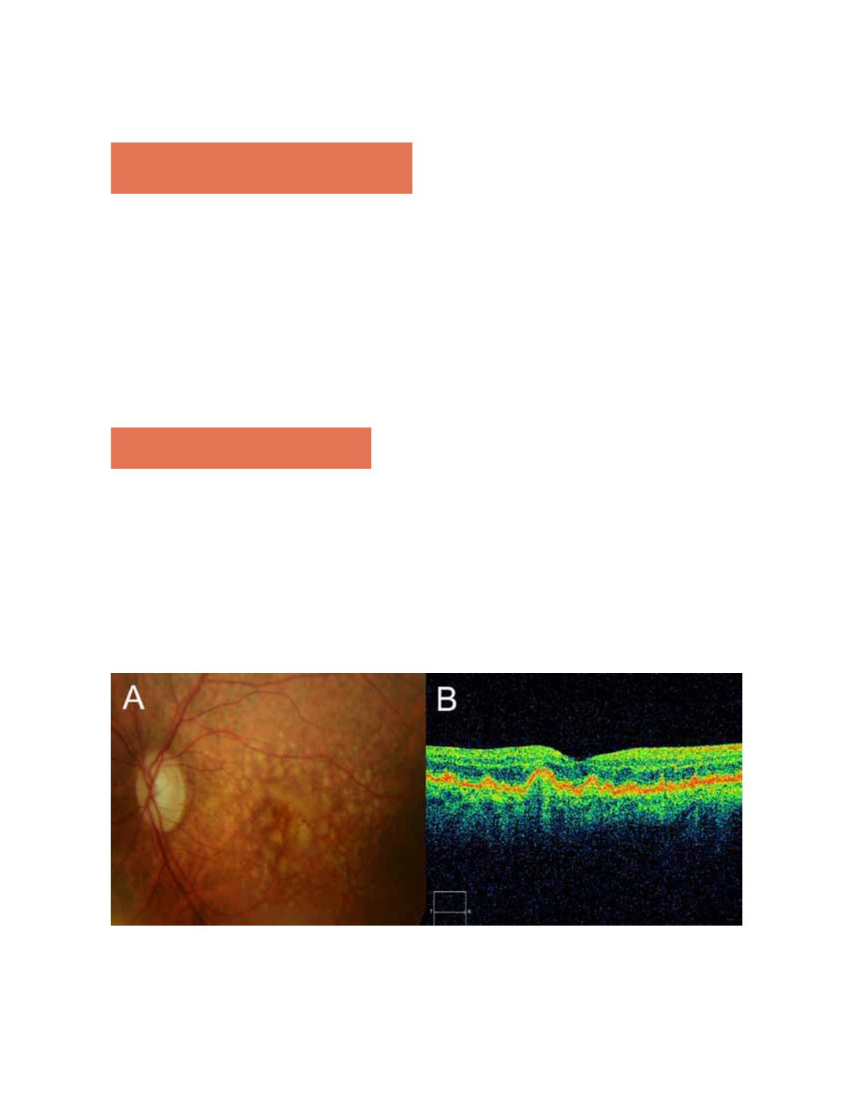
Figure 16.
(A) Central soft drusen may be seen in the retinography. (B) Drusen are observed as undulations and elevations of
RPE hyperreflective band with less reflective material below them.


