242
11. Time-Domain vs. Spectral-Domain OCT
in AMD
The new spectral-domain (SD) OCT devices include a
spectrometer in the receiver that analyzes the spectrum
of reflected light on the retina and transforms it into
information about the depth of the structures accord-
ing to the Fourier principle. This technology eliminates
the need to mechanically move the reference arm with
the consequent increase in the speed with which images
are received and axial resolution in time-domain (TD)
OCT.
30
In addition to greater speed in capturing images
and higher definition, the algorithms used by SD-OCT
differ from those of TD-OCT, and the retinal thickness
measurements are not comparable between the two.
While TD-OCT determines the total retinal thickness
by measuring the distance from the internal limiting
membrane to the highest hyperreflective band, i.e., that
combining the inner and outer segments of the photore-
ceptors, SD-OCT set this threshold in the RPE hyper-
reflective band, so the retinal thickness values are higher
than those obtained by TD-OCT (Figure 13).
31,32
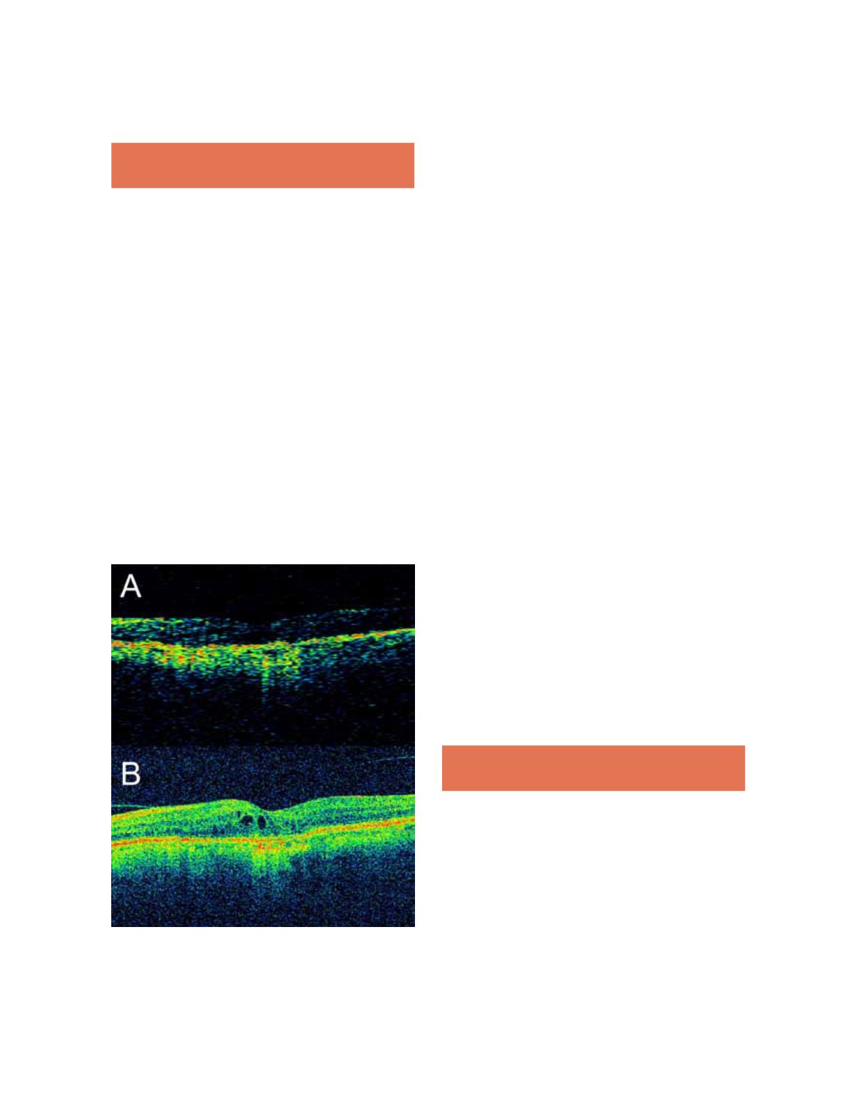
Figure 13.
SD- vs. TD-OCT. A, TD-OCT (Stratus). B, SD-
OCT (Cirrus). Foveal cysts are barely distinguishable with
the TD-OCT but are clearly identified by SD-OCT.
These differences justify the results of studies that compared
the two devices in patients with wet AMD. Thus, Mylonas
and colleagues found that in a number of patients with wet
AMD, the retinal thickness measurements obtained by
three SD-OCT devices were higher than those obtained by
TD-OCT instruments. The authors also emphasized the
importance of segmentation analysis as the main source of
errors with both devices.
33
The effect of this parameter had
already been studied widely for Stratus OCT (Carl Zeiss
Meditec Inc., Dublin, CA), and the proposed solution to
avoid these errors was the manual correction of segmenta-
tion lines in each image, especially in clinical trials of neo-
vascular AMD, in which the difficulty establishing the lim-
its in cases with subretinal and intraretinal fluid was auto-
matically higher.
22,34,35
To assess the incidence of errors at
this level in the SD-OCT apparatus, Krebs and colleagues
assessed 104 patients with neovascular AMD with both
TD-OCT and SD-OCT and analyzed the position of the
lines drawn automatically by segmentation analysis in each
case.
36
The results showed differences between the devices,
with TD-OCT committing errors in 69.2% of cases and
SD-OCT in 25%. These data suggested that SD-OCT
makes fewer errors in automatic segmentation analysis, and
these can be corrected manually identical to TD-OCT and
therefore constitute a marked improvement in the main
source of erroneous measurements. Finally, some studies
have compared both devices in a series of patients treated
with antiangiogenic agents. Sayanagi and colleagues com-
pared tomographic findings with TD-OCT and SD-OCT
in 58 patients with wet AMD treated with ranibizumab
(Lucentis, Novartis) and concluded that SD-OCT is better
than the TD linear mode B and mode 3D cube for detect-
ing intraretinal cysts and intraretinal and subretinal fluid
or fluid under the RPE, making it a more effective tool for
managing these patients.
37
12. Correlation with Other Techniques in
AMD
OCT retinal studies in patients with AMD can com-
plement information obtained with other conventional
examinations, such as angiography, or other more modern
techniques such as autofluorescence. We discuss combin-
ing these techniques in recent studies.
OCT and Angiography
Because of the widespread use of OCT, the use of FA has
dropped to second place in routine consultations, but it is


