238
8. Polypoidal Choroidopathy
Polypoidal choroidopathy is considered a variant of neo-
vascular membranes with serous or hemorrhagic detach-
ment of the retina and RPE in the posterior pole. The
pathology is seen on FA and especially by videoangiog-
raphy with green indocyanine green (ICG). Both tests
show dilated vessels in the choroid layer with the char-
acteristic presence of polypoidal structures which usually
occur at the termini of vessels at the edge of the vascular
network. (Figure 10).
8,24-26
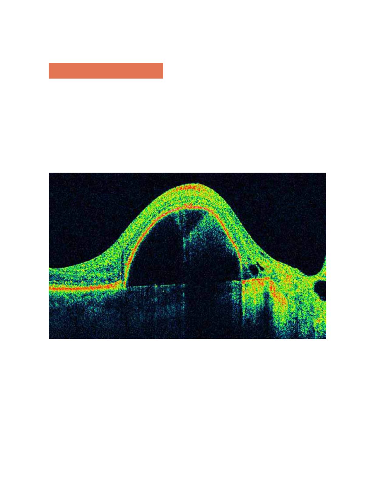
Figure 10.
OCT shows small cup-shaped RPE elevations and choroidal angiomatous lesions. Hemorrhagic and serous detach-
ments of the retina and the RPE also can be seen.


